
A Projection Of A Lizard's Pineal Organ
Lampreys have two parietal eyes one that developed from the parapineal organ and the other from the pineal organ. In avian species possessing a relatively conspicuous afferent projection such as Passeriformes Nycticorax and Milvus terminals of catecholamine-containing nerve fibers were observed exclusively in the interfollicular and perivascular.

Pin On The Man And L Uomo E
The zebrafish pineal organ is a photoreceptive structure containing two main neuronal populations photoreceptors and projections neurons.

A projection of a lizard's pineal organ. The pineal gland is located in the epithalamus near the center of the. A projection of a lizards pineal organ has an undeveloped retina and senses changes in light. The pineal complex has been retained in all vertebrates except crocodilians and a few mammals whereas the parietal eye has been lost in almost all clades except the lizards and tuataras.
Pineal Complex of Reptiles. Although not shown for lizards the achromatic units of photo- receptive pineal organs in many other groups exhibit a linear relationship with the logarithm of the luminance which is independent of light adaptation Dodt 1973. It is formed by two structures.
The pineal organ and the parietal eye. The pineal organ of fish is a photosensitive structure that receives light information from the environment and transduces it into hormonal rhythmic melatonin secretion and neural efferent projectionsneurotransmitters signals. It is formed by two structures.
Furthermore scattered labeled nerve fibers occur in different portions of the pineal stalk. The pineal gland in the adult brain is a midline structure that exhibits highly variable shapes among vertebrate species and is often associated with an accessory organ such as the parapineal organ in lampreys and fishes the frontal organ in amphibians and the parietal eye in lacertilian reptiles. Three-dimensional reconstruction of the distributional patterns of both types of neural projections was performed for the pineal organ of every avian species examined.
A DiI-tracing study of the neural connections of the pineal organ in two elasmobranchs Scyliorhinus canicula and Raja montagui suggests a pineal projection to the midbrain GnRH-immunoreactive nucleus. Pineal efferent projections have been traced in the brain of the goldfish Carassius auratus by administration of a concentrated solution of horseradish peroxidase onto the pineal organ. The comparative anatomy of median and lateral eyes with special reference to the origin of the pineal body.
Mandado M Molist P Anadón R Yáñez. After different survival times fish were sacrificed and the administered peroxidase was revealed by immunocytochemistry on paraffin sections using an anti. In this study we focused on this neural output.
The distribution of the central neural connections of the pineal organ of the larval sea lamprey was investigated by means of anterograde and retrograde tracing with the fluorescent lipophilic dye. Pinealofugal projections are well developed in larvae extending from the. A projection of a lizards pineal organ has an undeveloped retina and senses changes in light.
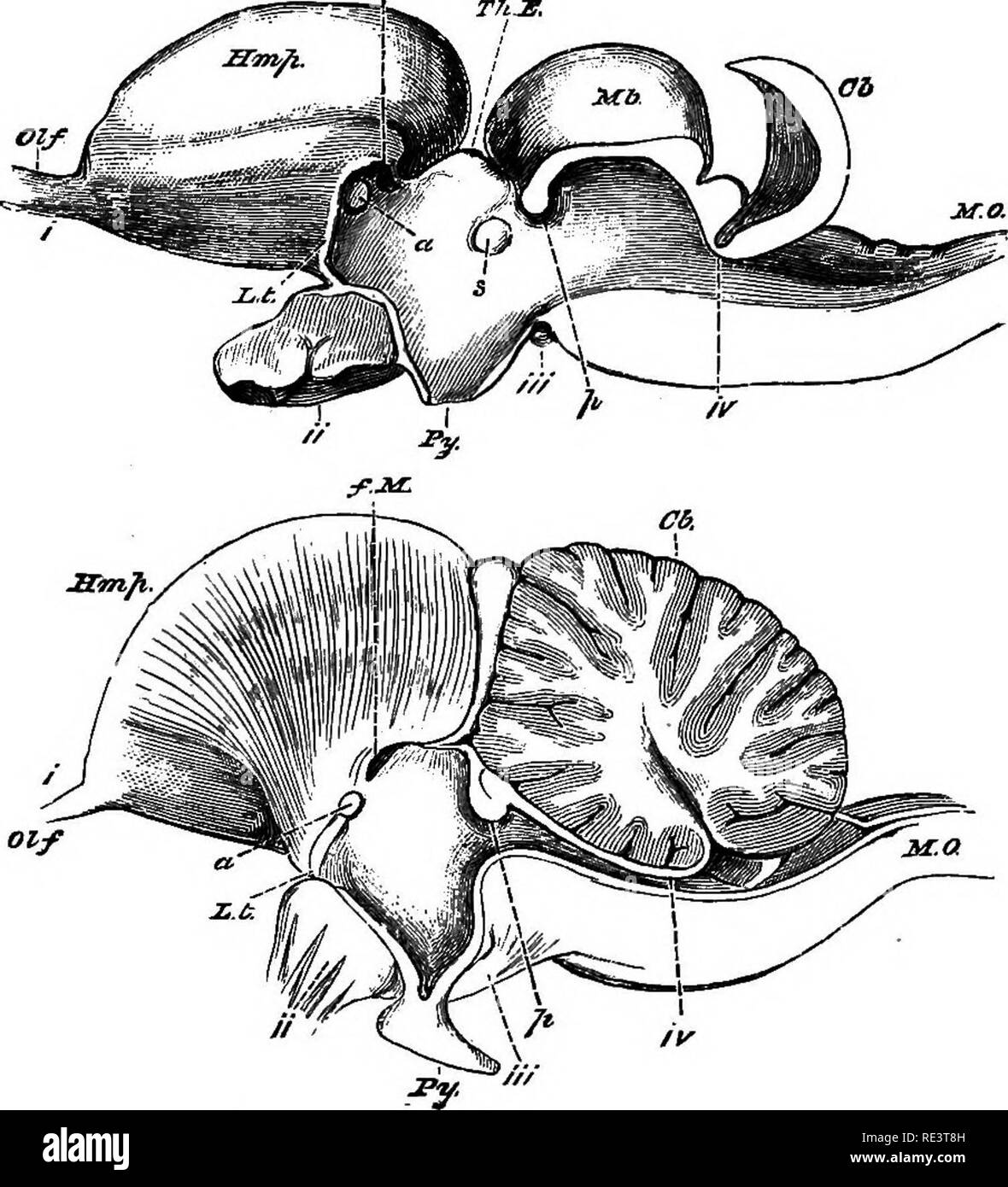
3 is largely variable in its organization. These are one behind the other in the centre of the upper surface of the braincase. They can be regarded either as processes of intrapineal neurons or projections of pinealopetal axons originating from central neurons.
DiI 11-dioctadecyl 3333-tetramethylindocarbocyanine perchlorate. The pineal complex of reptiles Fig. A population of nerve cells and their pinealofugal afferent fiber systems were stained by means of the acetylcholinesterase method while catecholamine-containing pinealopetal efferent fibers were demonstrated with the use of the glyoxylic acid method.
Some reptiles can changed color because of specialized structures called A. Central connections of the pineal organ of the three-spined stickleback Gasterosteus aculeatus L were studied by the use of horseradish peroxidase HRP for anterograde tracing. What Is this projection called.
The parietal eye also called the third eye and the pineal organ proper Gundy and. The innervation of the pineal organ was studied in 26 avian species under particular consideration of comparative aspects. HRP-labeled nerve fibers are observed in the distal division end-piece of the pineal organ.
The pineal gland is present in Chelonia Squamata and Rhynchocephalia but not in Crocodilia. The pineal complex of reptiles is a morphologically and functionally con-nected set of organs that originates as an evagination of the roof of the dien-cephalon. The pineal gland conarium or epiphysis cerebri is a small endocrine gland in the brain of most vertebratesThe pineal gland produces melatonin a serotonin-derived hormone which modulates sleep patterns in both circadian and seasonal cyclesThe shape of the gland resembles a pine cone from which it derived its name.
The pineal complex of reptiles is a morphologically and functionally connected set of organs that originates as an evagination of the roof of the diencephalon. Gladstone Reginald John 1865-. The minor ventrally directed projection could be followed through the periventricular.
Notably the parietal eye has been lost in snakes which evolved from a common ancestor to lizards. The dorsal part of this projection gave rise to terminals in the deep pineal gland and pineal stalk whereas the caudal part of this projection sent terminating fibers into the area postrema. The intracranial pineal organ of lizards may possess characteristics better suited to a dosimetric f unction.
But in Calotes Sphenodon and many others the parietal organ stays quite separate from the brain Fig. In many reptiles a cord of tissue connects the parietal organ with the pineal body. The pineal tract terminates bilaterally in the habenular nuclei area praetectalis dorsal and ventral thalamic nuclei periventricular hypothalamus and dorsal tegmentum.
What is this projection called. Because lampreys are among the most primitive of all living vertebrates it is possible that this was the original condition among vertebrates and may have allowed bottom-dwelling species to sense threats from. In more than half of the lizard species the pineal complex consists of two separate components.
So why do lizards still have the parietal eye. In addition to PhRs the pineal gland contains projection neurons PNs 20 cells at 48 hpf which project to the ventral diencephalon Wilson and Easter 1991 and a population of AgRP2 cells that do not express neuronal markers nor exhibit a neuronal morphology and share molecular characteristics with retinal-pigment epithelium cells Shainer et al 2017 Shainer et al 2019. Here we describe a subpopulation of projection neurons that expresses the melanopsin gene opn4xa.
And a description of the human pineal organ considered from the clinical and surgical standpoints. Because of the eyelike structure and on account of its connection with the roof of diencephalon or with the pineal body in many reptiles the. This new pineal cell type that displays characteristics of b.
Pineal Photoreceptors Use Two Signalling Modes Neural And Download Scientific Diagram
![]()
Pineal Gland Black And White Stock Photos Images Alamy

The Only Known Jawed Vertebrate With Four Eyes And The Bauplan Of The Pineal Complex Current Biology
2
Pineal Gland Black And White Stock Photos Images Alamy
![]()
Pdf The Circadian System Of Reptiles A Multioscillatory And Multiphotoreceptive System

Retinal And Pineal Nerve Projections In The Brain Of Sh Some Of The Download Scientific Diagram

The Pineal Complex Of Sauropsidians Excluding Crocodiles And Its Download Scientific Diagram

Parietal Eye An Overview Sciencedirect Topics

The Only Known Jawed Vertebrate With Four Eyes And The Bauplan Of The Pineal Complex Current Biology

The Pineal Organ Of Vertebrates In The Course Of Evolution In Download Scientific Diagram

Life Is Short But Snakes Are Long Do Snakes Have A Third Eye

Retinal And Pineal Nerve Projections In The Brain Of Sh Some Of The Download Scientific Diagram

Microtomographic Sections And Reconstruction Of The Green Lizard Skull Download Scientific Diagram
Posting Komentar untuk "A Projection Of A Lizard's Pineal Organ"