
Scapular Y Projection
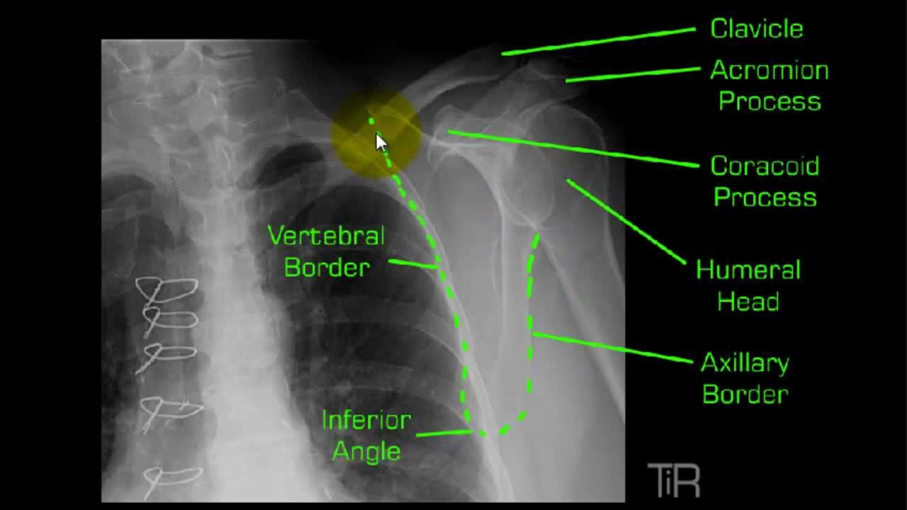
Scapular Y PA OBLIQUE PROJECTION RAO or LAO position Thi projection described by Rubin Gray and Green obtained its name as a result of the appearance of the scapula. Y-shaped intersection of the scapular body acromion pro-cess and coracoid process.

Pin On Xray
D Ferlic collimator-mounted filter used for AP and PA oblique scapular Y projections of shoulder.

Scapular y projection. If the anatomy and potential pathology are understood the radiography will be more meaningful. In order to reduce patient exposure this radiograph should not be repeated. Which projection of the upper limb should be taken to demonstrate a fracture of the proximal humerus when the arm.
The scapular Y projection is useful in evaluating anterior and posterior shoulder dislocations. To determine whether new positioning landmarks can help radiographers position the lateral scapula and Y projections more accurately. The acromioncoracoid process make up the upper legs of the Y the body makes up the lower leg of the Y.
This can be evaluated from this radiograph in spite of the poor positioning and exposure technique. This view is taken with the patient turned as for the Y projection and the cassette perpendicular to the body of the scapula and parallel to glenoid fossa. The glenoid fossa which forms the confluence of the Y being en face to the central x-ray beam and covered by the humeral head is usually not well seen on this projection.
The X-ray is taken from a mediolateral projection along the axis of the scapular spine with X-ray beam angled 1015 craniocaudally and centred on the acromioclavicular joint. The main purpose for the scapular Y projection is to evaluate the shoulder for dislocation. The humeral head should appear superimposed over the base of the Y if the humerus is not dislocated.
And the body the vertical stem. For the PA oblique projection scapular Y of the shoulder the body is rotated so the midcoronal plane MCP is how many degrees from the IR. Dislocations-to determine if anterior or posteriorly dislocated.
The proper name method for the AP oblique projection is the. Y-Scapula Projection Positioning Anatomy and Correction of the Malpositioned Radiograph AP Shoulder View Simulation. The thin body of the scapula should be seen on end without rib superimposition.
The acromion and coracoid processes should appear as nearly symmetric upper limbs of the Y. About Press Copyright Contact us Creators Advertise Developers Terms Privacy Policy Safety How YouTube works Test new features Press Copyright Contact us Creators. PA oblique projection of the shoulder scapular Y is performed to evaluate.
How much CR angulation should be used for a scapular Y projection. The ____ oblique scapular Y lateral projection of the scapula is taken with the patient in a anterior oblique position with the upper body rotated until the scapula is separated from the rib cage in a true lateral projection. Positioning for the scapular Y projection of the shoulder joint.
The humeral head positioned anterior to the glenoid fossa beneath the coracoid. The humerus superimposed over the scapular body. As much as the Y is for shoulder dislocations almost of doctors also want to check for supra-spinaious impingement.
Anatomy The acromion and coracoid form a Y or peace sign shape with the body of the scapula. Information from comparing the axillary and scapular Y-views demonstrates no significant difference in detecting abnormalities. The thin body of the scapula should be seen on end without rib superimposition.
E Ferlic collimator-mounted filter used for lateral projections of cervicothoracic region swimmers technique and axiolateral projections Danelius-Miller method of hip. A trauma lateral scapula projection is completely different to a Neers outlet projection both in its technique and objectives. The humeral head should be centeredovertheglenoidfossaThisviewcanbeveryhelpful in the setting of acute trauma to evaluate for anterior or posterior dislocation as the patient can be imaged with little or no movement of the arm and the projection obtains a.
Where is the CR centered for a transthoracic lateral projection. PA oblique projection of the shoulder scapular Y is performed to evaluate. Superimposition of the medial and lateral scapular borders.
A PA oblique scapular Y shoulder projection that shows accurate positioning of a patient with an anterior dislocation demonstrates. Radiographic Anatomy of a True Lateral Y-Scapula Projection. Currently used positioning landmarks for the lateral scapula and Y projections often yield inconsistent results and lead to repeats.
Level of the surgical neck. The scapula should be clearly demonstrated in the lateral profile. On the other axis if you see a the body of your scapula being projected between the top of your Y structure this is almost always cause for repeat.
This projection obtains its name as a result of the appearance of the scapula. The scapular spine its posterior arm. Clinicians highly recommend this view for the minimal shoulder motion necessary to obtain it 8.
For a scapular Y projection of the right shoulder which two of the following are acceptable options for positioning. Males have more sharply curved clavicles than females. F Ferlic collimator-mounted filter for AP axial projections of foot.
No CR angle should be used. The acromion and coracoid processes should appear as nearly symmetric upper limbs of the Y. What projection should be performed using a breathing technique.
The body of the scapula forms the vertical component of the Y and the acromion and. In this projection the anteriorly projecting coracoid process represents the anterior arm of the Y. The body of the scapula forms the vertical component of the Y while the acromion and coracoid processes form the upper limbs of the letter.

Shoulder Pectoral Girdle Shoulder Girdle Radiography Human Anatomy Glenohumeral Joint Shoulder Joint Acr Medical Anatomy Radiology Medical Knowledge

Pin On Surgery

Anatomy Vertebral Column Umls C1962945 Anterior Posterior Full Length View Of The Spine Lateral Full Length View O Radiology Anatomy Thoracic Vertebrae

Pin On Radtech
Pin On Radiology

Shoulder Dislocation Anterior Posterior Y View Scapular Xr Xray Shoulder Dislocation Radiology Imaging Radiology

Pin On For Radiology School

Pin On Xray

Pin On Retro Scientiarum

Pin On Xray

Boning Up On Humerus Clavicle And Ac Joint Positioning Diagnostic Imaging Radiology Radiology Student

Pin On Xray

Pin On Radiographic Images
Y View Shoulder Mp4 Radiology Nursing Radiology Schools Anatomy
Posting Komentar untuk "Scapular Y Projection"